






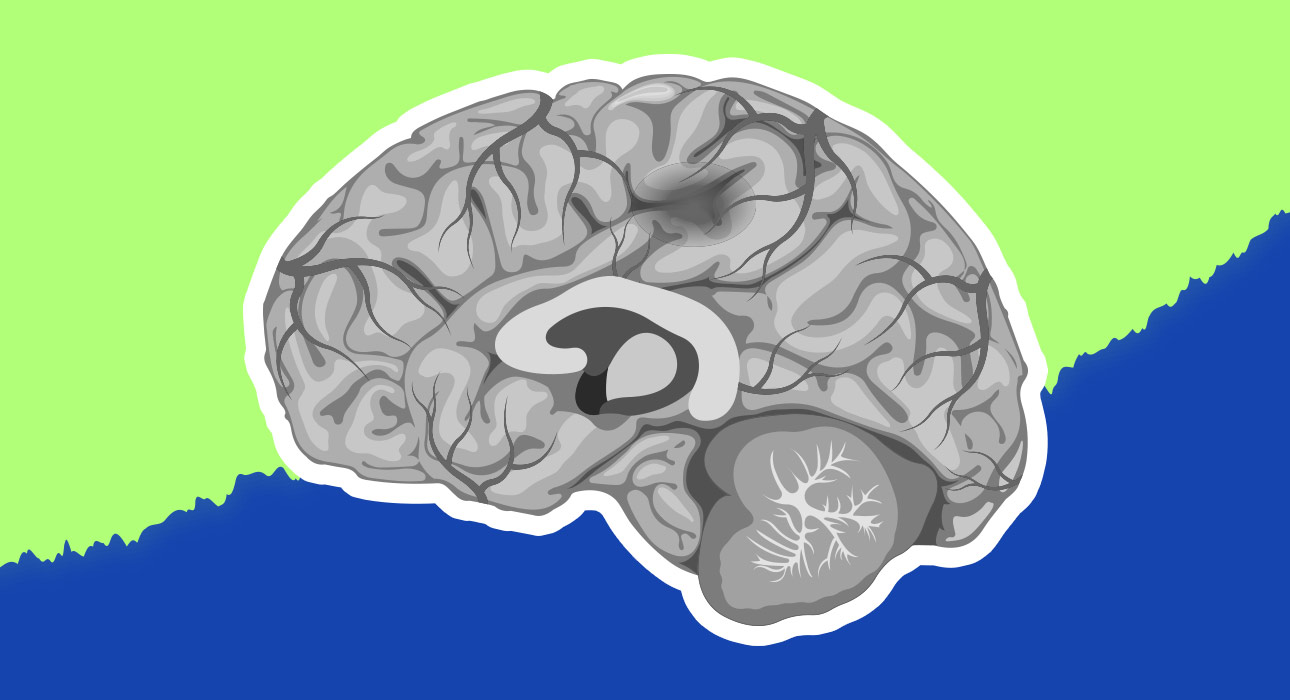
A stroke impacts about 15 million people worldwide, and out of those, about 5 million people have permanent damage (World Stroke Organisation, 2022). During a stroke, blood flow to part of the brain is suddenly blocked, either by a blockage in a blood vessel (ischaemic stroke, about 87 per cent of strokes) or by a burst blood vessel that leaks into the brain (haemorrhagic stroke, less common).
Brain cells require oxygen and glucose to survive and will die within minutes without them, which may cause permanent damage to motor function, speech, memory and cognition. The medical dogma for decades was that this damage was mostly permanent; loss of brain tissue meant loss of function. In the last three decades, however, neuroscience has shown that this is not the case. The article looks at recent scientific research into the brain’s adaptive capabilities after stroke, the factors that can facilitate or inhibit brain recovery, and how rehabilitation can help in brain plasticity to maximise restoration of function.
Read More: Most Powerful Ways Neuroplasticity Changes the Brain
Neuroplasticity: The Brain as a Plastic Organ
Neuroplasticity is the potential for the brain to adapt and restructure its abilities and functionality in response to experience, learning, and injury. It is not one but a family of related biological processes that take place on various time and length scales. Several different types of neuroplasticity come into play following stroke (Cramer et al., 2011).
1. Resolution of Diaschisis
The quickest process is the resolution of diaschisis- the temporary suppression or shutdown of neural activity in regions of the brain far from the site of the stroke, but connected to it via neural networks. Imagine a power failure in one section of the city, which leads to a reduction in the light in other sections of the city, but that does not cause them any harm. The swelling and inflammation of the damaged area will ease, and many of these distant areas will start to work again. Hence the fact that some stroke survivors make considerable improvement in the days and weeks after their stroke before they even begin formal rehabilitation (Carrera & Tononi, 2014).
2. Synaptic Plasticity and Long-Term Potentiation
The most significant neuroplastic change at the cellular level is synaptic plasticity, or strengthening or weakening of connections between individual neurons. This rule is sometimes referred to as “neurons that fire, wire together”. Repeated activation of a neural pathway through practice and experience strengthens and makes the pathways and synapses within it more efficient– this is called long-term potentiation (LTP).
3. Neuroplasticity in Stroke Rehabilitation
This is the biological mechanism underlying learning and recovery from stroke. With repeated engagement of the impaired function in a targeted activity, rehabilitation activates LTP in the remaining neural circuits, and over time, alternative circuits take over the function of the damaged circuits. Another way is through “cortical remapping”- a process whereby other areas of the brain assume a function once performed by the missing tissue.
4. Cortical Remapping After Stroke
Research with the imaging technique fMRI (functional MRI), which measures brain activity by identifying changes in blood flow, has demonstrated that after stroke, functions like hand movement or speech may move to neighbouring regions of the same hemisphere or to corresponding regions in the other hemisphere. This remapping does not happen automatically; it must be repeated and intentionally used on the involved function to facilitate the reorganisation (Nudo, 2013).
5. Neurogenesis and Stroke Recovery
Until recently, scientists believed that the adult human brain was incapable of forming new neurons. There is more recent research that shows limited neurogenesis (the formation of new neurons) occurs in certain brain areas, such as the hippocampus, the area that is central to the formation of new memories. There is evidence that stroke stimulates a certain amount of migration of neural precursor cells to damaged areas. But, in humans, it is not large enough to replace significant tissue loss directly, so it is rehabilitation, not regeneration, that is the most important process for meaningful functional recovery (Ohab et al., 2006).
Read More: The Role of Neuroplasticity in Personal Growth and Success
Factors that Shape the Brain’s Plasticity after Stroke
Even if the damage to the brain seems identical, not everyone who has a stroke recovers equally. Studies have revealed several biological, behavioural, and environmental factors that are critical in determining the amount of neuroplastic recovery an individual can experience.
1. Age
Younger brains tend to be more neuroplastic, i.e. more flexible in their structure and have more room for extensive remapping of the cortex. Older people, however, do not have to settle for this life-long disability. Even individuals in their 70s and 80s can have meaningful outcomes from intensive rehabilitation, and recovery outcome is not as well predicted by age as by the intensity of rehabilitation and extent of initial damage (Cramer et al., 2011).
2. Timing of Rehabilitation
This window of time following stroke is the time of highest neuroplastic potential, known as the critical window. In the first few weeks, the brain is in a very active state of being ready for reorganisation, partly due to increased growth factor activity. Early, intensive rehabilitation, which starts within days of onset of stroke, is associated with better outcomes compared with rehabilitation that is delayed by weeks or months (Langhorne et al., 2011). The discovery has changed the way stroke is treated, such as creating stroke units in hospitals where patients are offered rehabilitation as soon as they are in a state to receive it.
3. The Amount of Effort and Specificity of Practice
Learning is an active process, and repeated, challenging interaction with the very function that is impaired leads to neuroplasticity. Decades ago, research in animal models demonstrated that forced use of an impaired limb resulted in much more extensive cortical remapping than did passive assistance or substituting with the uncompromised limb. The finding, also observed in human studies, provides the scientific basis for constraint-induced movement therapy, which is discussed below (Taub et al., 2006).
4. Psychological and Emotional Issues
Depression, which occurs in about a third of stroke survivors, has a profound impact on neuroplastic recovery in several ways: by decreasing motivation; by decreasing involvement in rehabilitation; and through direct effects on the neurochemical environment in the recovering brain. Another chemical signal involved in synaptic plasticity is the neurotransmitter serotonin, which helps regulate emotions and feelings. Pharmacological and psychological treatments for post-stroke depression can improve rehabilitation outcomes beyond their effects on depressive symptoms alone (Robinson & Jorge, 2016). Family members can improve rehabilitation outcomes by actively supporting the individual’s participation in the rehabilitation process.
5. Cognitive Reserve
Cognitive reserve is the resistance of the brain, accumulated over a lifetime of education, socialisation, and mental stimulation, which helps it to withstand damage.
Individuals with high cognitive reserve may have greater potential for post-stroke neuroplastic reorganisation. This is because their brains may have more alternative pathways available to recruit when the primary pathways become damaged (Stern, 2009).
Read More: Rejuvenating Brain Neurons: A Breakthrough in Neuroplasticity and Mental Health
Rehabilitation Processes that tap into Neuroplasticity
The principle underlying modern stroke rehabilitation is that learning is an active process. It requires focused and intensive practice to stimulate neuroplasticity. These principles are reflected in evidence-based rehabilitation methods across the physical, cognitive, and speech domains.
1. Physical Rehabilitation
- One of the most extensively studied physical rehabilitation techniques is Constraint-Induced Movement Therapy (CIMT). Edward Taub and colleagues developed CIMT, which involves immobilising the unaffected limb, usually the arm and hand, while intensively training the affected limb for 2–3 weeks. This will force the brain to use the affected neural pathways instead of relying more heavily on the unaffected ones. Randomised controlled trials have demonstrated that CIMT yields significantly greater gains in arm and hand function than conventional therapy, and brain imaging has demonstrated the accompanying changes in representation of the affected arm in the brain (Taub et al., 2006).
- Another method is robotic-assisted therapy, which uses devices to guide the affected limb through desired movements while providing feedback that increases the number of repetitions and tasks performed (Huang & Krakauer, 2009), particularly in individuals with more severe impairments.
2. Cognitive Rehabilitation
Following stroke, there is a significant proportion of patients who have cognitive impairments, such as attention, memory, executive function (planning, organising and decision making) and processing speed, which affect independence and quality of life.
- Cognitive rehabilitation is a structured programme of exercises which target identified cognitive functions comprising computerised programmes, paper activities and practice with real-life activities. The systematic review by Cicerone et al. (2019) showed that specific cognitive rehabilitation programs for attention, memory and executive function deficits are effective following acquired brain injury such as stroke.
- Imagery involves patients picturing themselves performing a physical or mental exercise. It can also complement physical exercise by engaging similar neural pathways. This can enhance the benefits of traditional rehabilitation.
3. Rehabilitation of Speech and Language
Aphasia refers to the partial or total inability to speak, comprehend, read, or write language, and is one of the most bothersome effects on stroke survivors and their families (Engelter et al., 2006).
- Speech and Language Therapy: The evidence-based intervention is speech and language therapy (SLT), and there is a significant amount of research supporting the effectiveness of delivering SLT at an appropriate intensity.
- Constraint- Induced Aphasia Therapy: A forced use of language technique similar to CIMT is called Constraint-Induced Aphasia Therapy (CIAT), which asks patients to speak in a structured group setting while preventing them from using compensatory strategies like gestures or writing and has been found to be effective for enhancing naming, verbal fluency and communicative competence (Pulvermüller et al., 2001).
- (Melodic Intonation Therapy (MIT): MIT is a therapy that relies on melodic and rhythmic structures of singing to support speech output in patients with severe non-fluent aphasia, which is primarily supported by the right hemisphere, and has proven effective for patients with left hemisphere strokes (Norton et al., 2009).
Read More: Non-Directive Play Therapy: Healing Through the Language of Play
4. Non-invasive brain Stimulation
Transcranial Magnetic Stimulation (TMS) and transcranial direct current stimulation (tDCS) use magnetic or electrical fields to increase or decrease the excitability of targeted brain regions and promote cortical remapping, either by increasing activity in the damaged hemisphere or by reducing compensatory over-reliance on the unaffected hemisphere. A 2017 meta-analysis showed that tDCS + SLP was significantly more beneficial than SLP alone for aphasia (Elsner et al., 2017). These technologies are still mostly in the research and specialist clinical domains. However, they represent a promising front in the field of rehabilitation directed at neuroplasticity.
Read More: How Stroke Affects Speech Processing in the Brain
Conclusion
The science of the brain and how it changes after stroke has changed the understanding and hope of stroke recovery. The limited availability of high-intensity, evidence-based stroke rehabilitation means that many stroke survivors do not receive the level of therapy required. This limits their ability to optimise neuroplastic recovery. Stroke survivors also undergo a high range of depression, fatigue, and social isolation. At the same time, the period of greatest plasticity is reducing with time.
Thirty years ago, medical experts believed that little could be done after the first few months following a stroke. They also believed that most brain damage was largely permanent. Now, the results of this research are quite different. They suggest tremendous plasticity in the brain during recovery. The brain strengthens synaptic connections, remaps the cortex, and increasingly recruits new neural pathways.
Such a reorganisation is not automatic. It needs the very things that good rehabilitation offers. These include early, intensive, repetitive, task-specific practice of the impaired functions in a supportive and nurturing environment. This environment also seeks to include the psychological and social aspects of rehabilitation. Research on neuroplasticity highlights the brain’s potential: even after severe injury, the brain can continue to adapt and recover.
References +
- Carrera, E., & Tononi, G. (2014). Diaschisis: Past, present, future. Brain, 137(9), 2408–2422. https://doi.org/10.1093/brain/awu101
- Cicerone, K. D., Goldin, Y., Ganci, K., Rosenbaum, A., Wethe, J. V., Langenbahn, D. M., … Trexler, L. (2019). Evidence-based cognitive rehabilitation: Systematic review of the literature from 2009 through 2014. Archives of Physical Medicine and Rehabilitation, 100(8), 1515–1533. https://doi.org/10.1016/j.apmr.2019.02.011
- Cramer, S. C., Sur, M., Dobkin, B. H., O’Brien, C., Sanger, T. D., Trojanowski, J. Q., … Vinogradov, S. (2011). Harnessing neuroplasticity for clinical applications. Brain, 134(6), 1591–1609. https://doi.org/10.1093/brain/awr039
- Elsner, B., Kugler, J., Pohl, M., & Mehrholz, J. (2017). Transcranial direct current stimulation (tDCS) for improving aphasia in adults with aphasia after stroke. Cochrane Database of Systematic Reviews, 5, CD009760. https://doi.org/10.1002/14651858.CD009760.pub3
- Engelter, S. T., Gostynski, M., Papa, S., Frei, M., Born, C., Ajdacic-Gross, V., … Lyrer, P. A. (2006). Epidemiology of aphasia attributable to first ischemic stroke. Stroke, 37(6), 1379–1384. https://doi.org/10.1161/01.STR.0000221815.64093.8c
- Huang, V. S., & Krakauer, J. W. (2009). Robotic neurorehabilitation: A computational motor learning perspective. Journal of NeuroEngineering and Rehabilitation, 6, 5. https://doi.org/10.1186/1743-0003-6-5
- Langhorne, P., Bernhardt, J., & Kwakkel, G. (2011). Stroke rehabilitation. The Lancet, 377(9778), 1693–1702. https://doi.org/10.1016/S0140-6736(11)60325-5
- Norton, A., Zipse, L., Marchina, S., & Schlaug, G. (2009). Melodic intonation therapy: Shared insights on how it is done and why it might help. Annals of the New York Academy of Sciences, 1169(1), 431–436. https://doi.org/10.1111/j.1749-6632.2009.04859.x
- Nudo, R. J. (2013). Recovery after brain injury: Mechanisms and principles. Frontiers in Human Neuroscience, 7, 887. https://doi.org/10.3389/fnhum.2013.00887
- Ohab, J. J., Fleming, S., Blesch, A., & Carmichael, S. T. (2006). A neurovascular niche for neurogenesis after stroke. Journal of Neuroscience, 26(50), 13007–13016. https://doi.org/10.1523/JNEUROSCI.4323-06.2006
- Pulvermüller, F., Neininger, B., Elbert, T., Mohr, B., Rockstroh, B., Koebbel, P., & Taub, E. (2001). Constraint-induced therapy of chronic aphasia after stroke. Stroke, 32(7), 1621–1626. https://doi.org/10.1161/01.STR.32.7.1621
- Robinson, R. G., & Jorge, R. E. (2016). Post-stroke depression: A review. American Journal of Psychiatry, 173(3), 221–231. https://doi.org/10.1176/appi.ajp.2015.15030363
- Stern, Y. (2009). Cognitive reserve. Neuropsychologia, 47(10), 2015–2028. https://doi.org/10.1016/j.neuropsychologia.2009.03.004
- Taub E, Uswatte G, Elbert T. New treatments in neurorehabilitation founded on basic research. Nat Rev Neurosci. 2002 Mar;3(3):228-36. doi: 10.1038/nrn754. PMID: 11994754
- World Stroke Organisation. (2022). Global stroke fact sheet 2022. https://www.world-stroke.org/assets/downloads/WSO_Global_Stroke_Fact_Sheet.pdf



Leave feedback about this