







Consider the case of someone with a brain injury. While the person may observe the physical signs of this injury, another process is happening within the brain; this is where immune cells known as microglia react to the injury. The microglia continuously survey the brain, assist in removing injured brain cells, and aid the brain in its recovery process. But when their activation continues over time, the response can become problematic for the brain.
The human brain has been viewed as an organ that exists independent of the mechanisms that protect other organs in the body. This belief is now regarded as being wrong owing to studies done on the microglial cells. Microglial cells are the immune cells in the brain, which migrate into the brain during the process of brain development and remain in the brain for life. The cells keep watch for any harm within the brain. This article explores how microglia contribute to brain development, maintain brain health, respond to injury, and influence neurodegenerative and mental health conditions.
Read More: Examining Brain Immune Cells in Alzheimer’s Disease Progression
Origins and Development of Microglia
In contrast to other cells of the central nervous system, microglial cells do not have their origins in neural precursors. The fate-mapping studies have revealed that microglia are derived from yolk sac rudimentary macrophages and migrate into the brain at the stage when the blood-brain barrier has not yet formed; hence, microglia have been renewing locally throughout life without replacement from blood cells (Ginhoux et al., 2010).
Thus, this different origin makes microglia capable of functioning within the nervous system both as immune protectors and as neural circuit builders. In the early postnatal period, microglia perform the function of engulfing surplus and/or ineffective synaptic connections in a phenomenon called synaptic pruning, thus contributing to the development of neural connections necessary for normal cognitive and sensory development (Paolicelli et al., 2011).
Maintaining Brain Health: Surveillance and Homeostasis
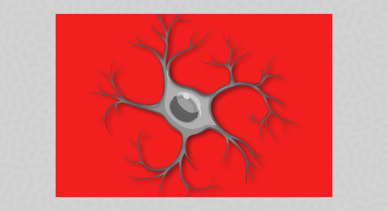
In the adult brain in a normal physiological state, microglia have a highly branched morphological structure, which was previously believed to be a quiescent form. The “resting” microglia observed under in vivo microscopy have been found to be continually active, as they keep extending and retracting their delicate processes in order to monitor their immediate surroundings multiple times every hour (Nimmerjahn et al., 2005).
Microglia are thus able to sense any alteration in the microenvironment and respond to local damage to ensure the maintenance of tissue homeostasis by removing waste products, dead cells, and misfolded proteins. Apart from their housekeeping roles, microglia secrete neurotrophic factors involved in the survival of neurons, synaptic plasticity, and adult neurogenesis (Salter & Stevens, 2017).
Response to injury
When the central nervous system gets hurt microglia are usually the cells to do something about it. If there is a problem like the blood-brain barrier gets broken the microglia near the problem will move fast to the damaged area. They do this within minutes. The microglia will then try to fix the problem by closing it off and stopping things from getting worse. This happens because the microglia change what they are doing. Normally they patrol the area but when there is a problem, they stop doing that and focus on fixing it. They will gather around the damaged area.
Form a kind of barrier to keep things from getting worse. At the time they will also eat up the bad cells and clean up the area. If someone has a brain injury or a stroke the microglia can really help at first. They can stop the damage from spreading. Help the brain fix itself. If the problem does not go away the microglia can get stuck in a mode where they are always trying to fix things even when it is not helping. This can actually make things worse, so can microglia.
Microglial Activation and Neuroinflammation
In the event that the microglia come across foreign material, such as harmful cells or substances that have no business being inside the body, they transition from surveillance to active engagement in which they devour the dangerous items and signal to other cells. The microglia do this by changing into a shape that can eat up the bad things and send out these signals.
The microglia are very good at helping us when we are sick or hurt. They help to keep the things from spreading and start to fix the damage. Sometimes the microglia can get too excited and keep sending out these signals for too long. This can hurt the cells in our brain and make us even sicker. The microglia can send out signals like interleukin-1β and tumour necrosis factor alpha when they are active. These signals can be very helpful when we are sick. They can also be very bad for us if they keep being sent out for too long.
Microglia in Neurodegenerative Disorders
Microgliosis is understood to be a fundamental part of neurodegenerative conditions such as Alzheimer’s disease, Parkinson’s disease, and multiple sclerosis, not just a result of cell death (Heneka et al., 2015). Microglia cells in Alzheimer’s disease interact with amyloid-beta plaques and, importantly, may reinitiate the developmental synaptic pruning mechanism that was mentioned above.
Scientists working with mouse models found that complement proteins tag vulnerable synapses for phagocytosis by microglia cells long before the formation of plaques, thus causing synapse removal that correlates with cognitive impairments (Hong et al., 2016). Researchers have found that Alzheimer’s disease involves more than the accumulation of protein aggregates. Brain immune cells, including microglia, actively contribute to the breakdown of neural networks. Similar inflammatory processes involving microglia have also been linked to the development of Parkinson’s disease and amyotrophic lateral sclerosis (ALS).
Read More: B-Cell Depletion Enhances Protective Gut-Derived Cells in Multiple Sclerosis
Microglia and Mental Health
Microglia also play an important role in psychiatric conditions, beyond their involvement in neurodegenerative diseases. The scientific literature has accumulated sufficient evidence to demonstrate the link between chronic low-level neuroinflammation and major depressive disorders; it has led some authors to call depression partly microglial disease (Yirmiya et al., 2015). Animal studies have shown that stress can affect hippocampal microglia both structurally and functionally. These changes can suppress neurogenesis and contribute to depression-like behaviour. Researchers have also found that inhibiting microglia can reverse these effects (Kreisel et al., 2014).
Studies in humans have found increased levels of inflammatory mediators in both the blood and brain tissue of people with depression. Moreover, research has shown that an inflammatory challenge can cause depressive symptoms in otherwise healthy individuals. This suggests a possible connection between the immune response and depression (Miller & Raison, 2016). Researchers have also identified unique microglial profiles. These profiles can help distinguish between healthy and pathological conditions. In the future, they may also serve as biomarkers for certain psychiatric conditions (Butovsky et al., 2014).
Conclusion
Microglia have two important roles in the central nervous system. They help protect and support the brain. However, when their activity becomes abnormal, they can also contribute to the development of diseases. Microglia have a unique origin. They develop during the embryonic stage and can regenerate throughout life. Their ability to monitor the brain and remove unnecessary synaptic connections is important for healthy brain development and stability (Ginhoux et al., 2010; Paolicelli et al., 2011).
However, the same characteristics that help microglia protect and support the brain can also cause problems when their activity becomes dysregulated. This can lead to excessive inflammation in the brain. Prolonged inflammation may contribute to neurodegenerative and mental health conditions, including depression (Heneka et al., 2015; Hong et al., 2016; Yirmiya et al., 2015). As research continues, scientists are working to understand the molecular signals that regulate changes in microglial behaviour. This research has changed how scientists view microglia. Scientists no longer consider them cells that only play a peripheral role in brain function. Instead, researchers increasingly recognise microglia as important players in maintaining brain health and contributing to the development of brain disorders (Salter & Stevens, 2017).
References +
- Butovsky, O., Jedrychowski, M., Moore, C. et al. Identification of a unique TGF-β–dependent molecular and functional signature in microglia. Nat Neurosci 17, 131–143 (2014). https://doi.org/10.1038/nn.3599
- Ginhoux, F., Greter, M., Leboeuf, M., Nandi, S., See, P., Gokhan, S., … Merad, M. (2010). Fate mapping analysis reveals that adult microglia derive from primitive macrophages. Science, 330(6005), 841–845. https://doi.org/10.1126/science.1194637
- Heneka, M. T., Carson, M. J., El Khoury, J., Landreth, G. E., Brosseron, F., Feinstein, D. L., … Kummer, M. P. (2015). Neuroinflammation in Alzheimer’s disease. The Lancet Neurology, 14(4), 388–405. https://doi.org/10.1016/S1474-4422(15)70016-5
- Hong, S., Beja-Glasser, V. F., Nfonoyim, B. M., Frouin, A., Li, S., Ramakrishnan, S., … Stevens, B. (2016). Complement and microglia mediate early synapse loss in Alzheimer mouse models. Science, 352(6286), 712–716. https://doi.org/10.1126/science.aad8373
- Kreisel, T., Frank, M., Licht, T. et al. Dynamic microglial alterations underlie stress-induced depressive-like behaviour and suppressed neurogenesis. Mol Psychiatry 19, 699–709 (2014). https://doi.org/10.1038/mp.2013.155
- Miller, A., Raison, C. The role of inflammation in depression: from evolutionary imperative to modern treatment target. Nat Rev Immunol 16, 22–34 (2016). https://doi.org/10.1038/nri.2015.5
- Nimmerjahn, A., Kirchhoff, F., & Helmchen, F. (2005). Resting microglial cells are highly dynamic surveillants of brain parenchyma in vivo. Science, 308(5726), 1314–1318. https://doi.org/10.1126/science.1110647
- Paolicelli, R. C., Bolasco, G., Pagani, F., Maggi, L., Scianni, M., Panzanelli, P., Giustetto, M., Ferreira, T. A., Guiducci, E., Dumas, L., Ragozzino, D., & Gross, C. T. (2011). Synaptic pruning by microglia is necessary for normal brain development. Science, 333(6048), 1456–1458. https://doi.org/10.1126/science.1202529
- Salter, M., Stevens, B. Microglia emerge as central players in brain disease. Nat Med 23, 1018– 1027 (2017). https://doi.org/10.1038/nm.4397
- Yirmiya, R., Rimmerman, N., & Reshef, R. (2015). Depression as a microglial disease. Trends in Neurosciences, 38(10), 637–658. https://doi.org/10.1016/j.tins.2015.08.001





Leave feedback about this