


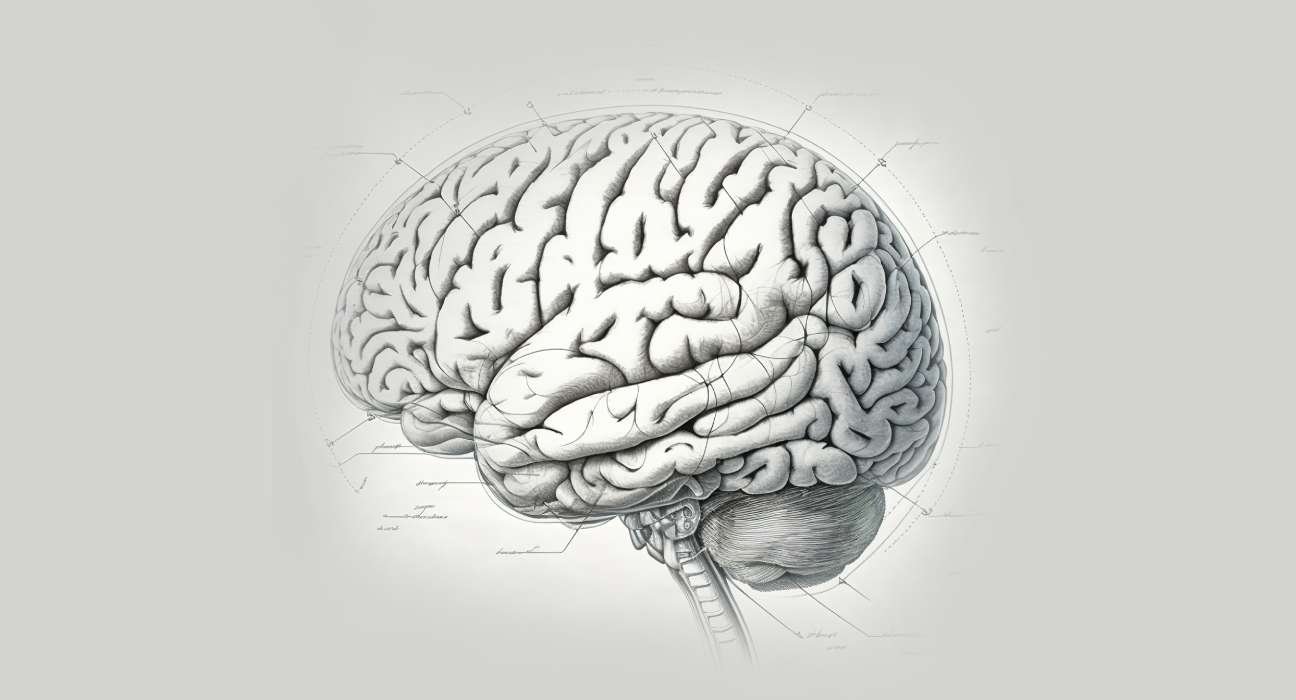
The cerebral cortex, or gray matter, is known as the outermost layer of nerve cell tissue in your brain. Several folds and grooves give it a wrinkly appearance. Memory, cognition, learning, reasoning, problem-solving, emotions, consciousness, and sense-related activities are all significantly influenced by your cerebral cortex. Also the brain’s outermost layer is called the cerebral cortex. Due to the numerous folds on its surface, it appears wrinkled. The folds are made up of several elevated regions called gyri and deep grooves called sulci. These folds increase the cerebral cortex’s surface area, which enables more nerve cells to process vast amounts of information. About half of brain’s overall mass is composed of your cerebral cortex.
Between 14 billion to 16 billion nerve cells are distributed among the six layers of your cerebral cortex. Its thickness is between two and four millimeters (mm; 0.08 and 0.16 inches). The right and left hemispheres of the brain are similarly divided, and they are separated by a sizable sulcus known as the medial longitudinal fissure. To enable communication and the formation of new connections between the two hemispheres of the cerebral cortex, bundles of nerve fibers known as the corpus callosum join them.
The lobes, which are separated according to the positions of gyri and sulci, are used by the cerebral cortex to regulate a wide range of functions. The frontal, temporal, parietal, and occipital lobes are the names given to these lobes. Processing of various kinds of information is the responsibility of each of these lobes. Language, memory, logic, cognition, learning, decision-making, emotion, intelligence, and personality are among the higher-order cognitive functions of the human brain that are collectively controlled by the cerebral cortex.
Also Read: Unlock Your Brain’s Power: Enhance Memory and Cognitive Performance
Why Is The Cerebral Cortex Known As Grey Matter?
Your brain’s outer layer, known as gray matter, is made up of nerve cell bodies, including dendrites, which are the tips of nerves. The portion of a nerve cell called a dendrite is responsible for receiving chemical messages from neighboring cells. Your cerebral cortex appears gray because the myelin, a fatty coating layer, is absent from that area of the nerve.
The lengthy, myelin-wrapped central segments of nerve cells, or axons, make up the bundles that make up the white matter in your brain. The tissue’s pale hue is attributed to myelin.
Difference Between Cerebrum And Cerebral Cortex
- The cerebral cortex is the outer layer of the cerebrum, which is the biggest portion of the brain.
- Gray matter, which consists of cell bodies and dendrites, covers the interior white matter in the cerebral cortex because the cerebrum is composed of nerve fibers and cell bodies, as well as gray and white matter.
- There are two hemispheres in the cerebrum. The frontal, parietal, temporal, and occipital lobes are the four lobes that make up the cerebral cortex.
- The cerebrum’s primary job is to regulate the body’s voluntary muscular motions. Consciousness is mostly mediated by the cerebral cortex
Functions
1. Frontal lobe
Behind your forehead, at the front of your brain, is the frontal lobe. Your frontal lobe performs the following tasks:
- Making decisions and resolving issues.
- Deliberate mental processes.
- Attention
- Behavioral and emotional restraint.
- Production of speech.
- Intelligence
- Motion of the body.

The motor cortex, prefrontal cortex, and Broca’s area are three noteworthy regions located within this lobe. Your body’s mobility is controlled by the motor cortex. Thinking and solving problems are examples of “executive functions” that are controlled by prefrontal cortex. It oversees and guides other brain regions as well. The portion of your frontal brain associated with producing speech is called Broca’s area.
Also Read: Brain waves for Bliss: Nurturing Well-being through Neuro-Harmony
2. Occipital Lobe
The brain’s occipital lobe is located toward the back. Among the occipital lobe’s functions are:
- Processing and interpretation of images.
- Gathering of visual information about color, motion, and orientation.
- Object and facial recognition.
- Perception of distance and depth.
- Visual world mapping.
3. Parietal lobe
Situated above the temporal lobe and between the frontal and occipital lobes is the parietal lobe. The parietal lobe performs the following tasks:
Processing of sensory data, including temperature, vibration, location, pressure, and pain. Both spatial manipulation and processing. This is the capacity to navigate around your home or town, or to know where you are in three dimensions. Within this lobe, the somatosensory cortex is one of the noteworthy areas. It gathers sensory data from every part of your body, or “feeling” data.
An illustration of how brain lobes cooperate is shown here:
The signal that tells the muscles in your hand and arm to reach toward a cup of soup on your kitchen table comes from the motor cortex in the frontal lobe of the brain. The parietal lobe’s somatosensory cortex evaluates the data you receive from touching the cup, including your assessment of its temperature. Your parietal lobe’s spatial processing enables you to hold the cup and move your hand perfectly between it and the table and other items in the environment.
Also Read: 10 habits that may damage your brain
4. Temporal Lobe
Your temporal lobe is situated beneath your parietal lobe and between your frontal and occipital lobes. Your temporal lobe performs the following tasks:
- Learning, speech production, and language Understanding.
- Recall.
- Listening.
- Nonverbal communication.
- Translation of sound into visual images.
Wernicke’s region is a noteworthy location inside this lobe. More recently, it was found that this region is involved in language comprehensiveness, connecting previously learnt sounds to voice tones and noises.
Different Areas Of Cerebral Cortex
Using a different perspective on the brain, some researchers categorize the cerebral cortex’s areas according to the three primary types of functions that they represent: sensory, motor, and association areas.
Sensory Area
Information from your environment and senses is sent to these parts of your cerebral cortex. Features consist of:
- Object recognition and the interpretation of visual data. The visual cortex, located in the occipital lobe, is responsible for processing these activities
- Evaluating your body’s touch, temperature, location, vibration, pressure, and pain signals. Your parietal lobe’s somatosensory cortex is responsible for processing these actions.
- Analyzing the data from the hearing. Your temporal lobe’s auditory cortex is responsible for processing this function.
- Analyzing flavor and taste. The gustatory cortex is a region of your frontal brain that handles these processes.
Motor Area
Your cerebral cortex’s motor regions are responsible for your voluntary muscular movements. The primary processing area for these functions is the frontal lobe. Among the functions are:
- Coordination of the movement of the muscles.
- Arranging for intricate movements.
- Empathy and imitation as learning tools.
Also Read: Brainstorming: A valuable technique for getting ideas
Association Area
These regions connect and enhance the complexity of functions, and they are dispersed throughout the four lobes. Among the functions are:
- Arranging and interpreting data from motor and sensory domains.
- Character traits and emotional self-control.
- Logic and awareness of space.
- Processing of memories.
- Think visually and remember things visually.
- Combine language, sound, and memories to create visual information.
3 main regions
There are three main regions that make up the cerebral cortex:
- Neocortex: Six layers make up the most recent evolutionary stage. Comprises over 90% of the cerebral cortex in humans. Higher order cognitive processes, including sensory perception, motor command synthesis, language, spatial reasoning, and conscious thought are handled by this system.
- Allocortex: The older, less than six-layered area is called the allocortex and Includes the hippocampal region and the olfactory cortex. Participates in memory and smell.
- Archicortex: The earliest portion, the archicortex, has just three cortical layers also, the hippocampus is formed. involved in both spatial navigation and memory.

Damage cerebral cortex
Any of the conditions can damage any area of the cerebral cortex: tumors, autoimmune illnesses, brain hemorrhage, or stroke. The location of the damage will determine the symptoms.
1) The Frontal lobe
The following are signs that your frontal lobe is damaged or injured:
- Problems with memory.
- A person’s personality can shift.
- Handling problems and making decisions.
- Issues with focus.
- Socially inappropriate behavior, emotional deficiency, and behavioral modifications.
- Incapacity to comprehend or articulate speech (aphasia).
- Speech difficulties (apraxia).
- Loss of muscle control, weakness, and paralysis on one side of the body (flaccid hemiplegia)
- Dementia
2) The Parietal lobe
- An inability to write (agraphia)
- Problems with math,
- Numbness,
- Disorientation,
- Loss of sensation.
- Aphasia.
- Apraxia.
- Poor hand-eye coordination,
- Loss of sensation,
- The inability to distinguish items solely by touch are all signs of damage to the parietal lobe.
Also Read: Why Is Exercise And Good Nutrition So Important For A Healthy Brain?
3) Damage to the frontal lobes
Your temporal lobe shows signs of damage if the following are observed:
- Difficulty hearing.
- Problems with memory.
- Inability to distinguish between objects and faces.
- Linguistic impairments, including Wernicke’s aphasia, and problems with language comprehension.
- Alzheimer’s disease, developmental dyslexia, and epileptic seizures are some conditions that can harm the temporal lobe.
4) The Occipital lobe
Symptoms of damage to your occipital lobe include:
- Difficulty perceiving more than one object at the same time.
- Trouble recognizing objects by sight.
- Colorblindness.
- Hallucinations involving vision.
- Total blindness.
Clinical Relevance
- An examination of the literature on the relationship between the frontal lobe and schizophrenia revealed that many patients’ frontal lobes differed from those of people without the disorder in terms of grey matter volumes and functional activity (Mubarik & Tohid, 2016), indicating a possible association between the frontal lobes and schizophrenia.
- Researchers have discovered that people with schizophrenia have less grey matter volume in their parietal lobes (Zhou et al., 2007). Structural abnormalities or atrophy in the parietal lobes may cause some of the symptoms of schizophrenia, implying that managing the illness could be facilitated by treating this area of the brain.
- According to Nasab et al. (2021) children with ADHD differed from neurotypical children in their emotional processing as evidenced by altered functional connectivity and decreased network efficiency in the frontal and occipital lobes while perceiving facial expressions.
- The temporal lobes’ attention deficits were found to be linked to developmental dyslexia by researchers.(Voldois et al., 2019)
- A review of research on brain imaging revealed that individuals with autism spectrum disorder and typically developing youngsters differed in the way their frontal lobes developed. Children with autism showed larger frontal lobe volumes between the ages of 2-4 years, indicating abnormal brain development in areas critical to cognitive abilities (Crucitti et al., 2022).
References+
● https://www.simplypsychology.org/what-is-the-cerebral-cortex.html
● https://my.clevelandclinic.org/health/articles/
● https://en.m.wikipedia.org/wiki/Cerebral_cortex
●https://www.google.com/sq=Children+with+ADHD+showed+altered+functional+connectivity+and+reduced+network+efficiency+in+the+frontal+and+occipital+lobes+https://www.simplypsychology.org/what-is-the-cerebral-cortex.html


Leave feedback about this