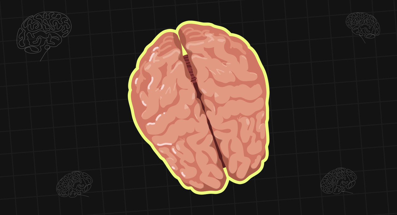
Imagine being shown an object that you can clearly see, yet being unable to say what it is. Now imagine something even stranger: you can still pick it up with one hand. This is not a thought experiment. Something remarkably similar was observed in patients whose corpus callosum, the major connection between the brain’s two halves, had been surgically cut. In one classic experiment, an object was briefly shown to the patient’s left visual field. The patient could not verbally identify what they had seen, but when asked to use their left hand to select the object, they could often choose it correctly.
What was happening? The object had not simply gone unnoticed. The visual information had reached the brain, but it had reached one half of the brain without being effectively communicated to the other half, which was responsible for producing speech. The patient’s experience revealed something that is normally hidden from us: although we experience ourselves as having one unified mind, the brain is made up of two cerebral hemispheres that constantly exchange information.
The main bridge between these hemispheres is the corpus callosum, a thick bundle of nerve fibres that connects the left and right sides of the cerebral cortex. It is the largest of the brain’s commissural fibre systems (van der Knaap & van der Ham, 2011). Understanding what happens when this connection is cut or when it fails to develop gives us a fascinating window into how the two hemispheres cooperate, what happens when they cannot communicate, and why the popular idea that people are simply “left-brained” or “right-brained” is far more complicated than it appears.
Read More: The Split-Brain Patients Who Changed Our Understanding of Consciousness
A Network, Not A Single Cable
The corpus callosum is made up of millions of myelinated nerve fibres which cross the midline of the brain and link up the corresponding areas in the two hemispheres (van der Knaap & van der Ham, 2011). It does not have a uniform structure; it is divided into five parts, namely the rostrum, genu, body, isthmus and splenium, and it is these regions that connect widely different areas of the cortex (Aboitiz et al., 1992; van der Knaap & van der Ham, 2011).
The fact that the organisation is like this shows that the various brain systems have to exchange different types of information. The splenium, which is at the back, plays a major role in transferring visual information between the two hemispheres, while the fibres that connect the motor areas help to co-ordinate movements involving both sides of the body (Hervé et al., 2006; Wahl & Ziemann, 2008).
The corpus callosum is therefore more accurately seen as a communication network that carries a number of different kinds of information rather than as a single telephone line. A result of this is that its function cannot be reduced to one single role; different areas serve different systems, and the effects of any disruption will depend on how much the fibres are affected (Aboitiz et al., 1992; van der Knaap & van der Ham, 2011).
Read More: Cerebral Cortex: The most important part of human brain
Specialised Hemispheres That Must Cooperate
Although the two halves of the brain are generally symmetrical, they are not functionally the same. There is lateralisation in some abilities, which means that one hemisphere is more involved than the other. For instance, language is usually more laterally organised in the left hemisphere, while certain aspects of visuospatial processing tend to involve the right hemisphere more (Gazzaniga, 2000).
Yet lateralisation does not mean that one hemisphere works by itself. Take the example of reading a map while you are travelling. In order to interpret the layout, visuospatial processing is required, language is needed to read the labels, memory has to be used to link the map with previous knowledge, and the motor systems must be employed to steer or to point in a certain direction.
These various functions are spread out over different networks rather than being confined to one side of the brain. The corpus callosum enables specialised systems to co-ordinate and communicate, so that the brain can combine the information rather than functioning as two separate brains (Bloom & Hynd, 2005; van der Knaap & van der Ham, 2011). A suitable analogy would be two colleagues who have different areas of expertise working on a common project. Although their skills are different, that is actually the point in favour of having them, but the project can only be completed if they share what they know.
From Sensation To Meaning
When we look at sensory information, the importance of communication between the two hemispheres becomes more apparent. For example, if an unknown object is placed in your left hand and your eyes are closed, the information from the left side of the body is mainly dealt with by the right hemisphere. Since recognition of the object will involve tactile, visual and conceptual information which is spread over various areas of the brain, the corpus callosum enables the information processed in one hemisphere to interact with the information that is represented in the other (van der Knaap & van der Ham, 2011).
The same principle holds true in the case of vision. Each half of the visual field is at first mainly processed by the opposite hemisphere, but there are fibres from the corpus callosum that link the visual areas on either side. The posterior part of the corpus callosum, especially the splenium, is involved in the communication between the various visual cortical areas and aids in the integration of visual processing across the midline (Hervé et al., 2006).
It is because of this integration that the visual world does not seem to be made up of two separate halves; the experience of a single, unbroken scene is due to an incredibly complex system of neural communication, a system which goes largely unnoticed unless it is disturbed (Hervé et al., 2006).
Read More: Predictive Processing: Is the Brain Really a Prediction Machine?
What Happens When Researchers Cut the Connection?
Researchers gain valuable insights into the function of the corpus callosum by studying patients who have undergone a corpus callosotomy, a surgical procedure that cuts part or all of this structure. Surgeons traditionally performed this procedure to treat severe epilepsy because it prevented seizure activity from spreading from one hemisphere to the other (van der Knaap & van der Ham, 2011).
Roger Sperry and Michael Gazzaniga extensively studied these patients through what became known as split-brain research, significantly advancing scientific understanding of hemispheric specialisation (Sperry, 1968; Gazzaniga, 2000). In normal day-to-day situations, these patients usually seemed completely normal and were able to speak, walk, recognise people and carry out a great many routine tasks. Gazzaniga (2000) found that carefully designed experiments could demonstrate situations in which one hemisphere could not access information presented to the other.
A typical example of this is the visual system. When an object briefly appears in the left visual field, the right hemisphere processes the visual information. Because the left hemisphere primarily controls language, a person may struggle to name an object even when the right hemisphere has successfully processed it. However, they can at times identify or select the object using their left hand, which is more directly under the control of the right hemisphere (Gazzaniga, 2000).
The short duration of the presentation is a key feature of the experiment. If the image had stayed on the screen, the participant could have moved their eyes so that both hemispheres could see it, and the effect would then have disappeared. It was the ability to control which hemisphere received the information that made these experiments useful (Gazzaniga, 2000).
Gazzaniga (2000) emphasised that neither hemisphere had become superior to the other; instead, the two hemispheres could no longer exchange information that they would normally share. The difference between an inability to process information and an inability to share it changed the way researchers considered the role of each hemisphere (Sperry, 1968; Gazzaniga, 2000).
Read More: 6 Mind-Blowing Neuroscience Facts About the Human Brain
More Than An Information Cable
Scientists view the corpus callosum as more than just a cable that transfers information between the hemispheres. Research indicates that communication through the corpus callosum can produce both excitatory and inhibitory effects. At times, the two hemispheres can enhance each other’s activity. At other times, they can regulate or suppress competing activity (Bloom & Hynd, 2005; van der Knaap & van der Ham, 2011).
A system which has two halves probably needs both of these functions since it must combine useful signals while at the same time preventing the two halves from interfering with each other. We can observe this in coordinated movements. For example, when playing the piano, typing, cutting vegetables or tying shoelaces, both hands may have to carry out different but accurately coordinated actions. The communication between the motor areas of the two hemispheres plays a role in this coordination (Wahl & Ziemann, 2008).
Recent reviews of bimanual coordination have also shown links between the structure of the corpus callosum and the ability to coordinate movements with both hands (Gooijers & Swinnen, 2014; Fling et al., 2023). The corpus callosum helps share information between the two hemispheres. It also supports the timing and coordination of activity between them (Wahl & Ziemann, 2008; Gooijers & Swinnen, 2014).
Read More: Thalamus: One of the Essential Parts of the Human Brain
When The Corpus Callosum Never Develops
Disconnection may also take place without having surgery. Agenesis of the corpus callosum happens when the structure does not develop properly before birth. Notably, people with agenesis do not simply show the same pattern as those who have had surgical disconnection (Siffredi et al., 2013; Brown & Paul, 2019). One explanation is that the developing brain can sometimes compensate for this absence. It does so by using alternative pathways and reorganising its networks (Siffredi et al., 2013; Brown & Paul, 2019).
As a result, outcomes vary greatly among individuals. Some show relatively typical intellectual functioning. Others experience difficulties with processing speed, complex reasoning, executive functions, social cognition, or the transfer of sensory-motor information (Siffredi et al., 2013; Brown & Paul, 2019; Wright & Booth, 2023). This variability is significant because it shows that a single brain structure rarely determines an ability on its own.
Development can also influence how different networks communicate (Brown & Paul, 2019; Wright & Booth, 2023). It also suggests that individuals without a corpus callosum do not share a single, predictable profile. Therefore, researchers approach these groups with caution (Siffredi et al., 2013; Wright & Booth, 2023).
Read More: Why Your Brain Keeps an Audio Replay: Echoic Memory
Two Hemispheres, One Experience
Split-brain studies have raised a question that goes beyond anatomy. If the two hemispheres can function separately, what creates the feeling of being a single person? In everyday life, the answer appears obvious, as people do have one continuous stream of experience, one set of intentions and one voice.
The corpus callosum contributes to this sense of unity by allowing sensory, motor, and cognitive systems to share information. It also helps these systems work together in a coordinated manner (Bloom & Hynd, 2005; van der Knaap & van der Ham, 2011). For example, reaching for a cup involves vision, movement, memory, and decision-making. However, these processes work together smoothly without conscious effort. The smoothness of the experience masks the extent of the cooperation needed between the specialised systems.
Studies of disconnection show that the smooth integration of brain functions depends on communication between different areas. This integration does not happen automatically (Sperry, 1968; Gazzaniga, 2000). These studies do not prove that a patient with a split brain becomes two separate individuals. The best way to interpret their experiences also remains a subject of debate. However, these findings show that the unity people take for granted has a physical basis. Gazzaniga (2000) also demonstrated that researchers can disrupt this unity in specific and measurable ways.
Read More: Know About Brain: Eliminating Myths, Correcting Facts
The Myth Of The “Left-Brained” Or “Right-Brained” Person
People often describe themselves or others as “left-brained” or “right-brained” to suggest a preference for logical thinking or creativity. These phrases associate logic and analytical thinking with one hemisphere and creativity with the other. They also imply that each person tends to favour one side of the brain. The brain lateralises certain functions, meaning that one hemisphere plays a more dominant role in performing them. However, Gazzaniga (2000) cautioned against the misleading idea that people primarily rely on either their left or right hemisphere.
Contemporary neuroscience places more emphasis on networks and interactions than on separate hemispheres. Although different functions may show a preference for one hemisphere, they still depend on communication throughout the brain. Studies into cross-hemispheric communication indicate that the interactions between the two hemispheres can affect how laterally specialised functions develop and function (Gazzaniga, 2000; Zaidel et al., 2003).
Handedness provides a helpful point of comparison. Most people have a preferred hand. However, they rarely use only that hand. Even when performing complex tasks, the preferred hand often relies on the other hand for support. Similarly, the brain is not a competition between its two hemispheres. Instead, it involves collaboration between specialised systems (Gazzaniga, 2000; Zaidel et al., 2003).
Read More: What Is DMI Test How Does It Measure Brain Capacity
Conclusion
The corpus callosum receives less attention than the frontal lobe, hippocampus, or amygdala. However, it plays a central role in helping the brain function as an integrated system. It allows sensory information to pass from one hemisphere to the other. It also helps coordinate different motor systems. Additionally, it supports cognitive integration. It also allows specialised brain areas to communicate despite performing different functions (Bloom & Hynd, 2005; van der Knaap & van der Ham, 2011; Wahl & Ziemann, 2008).
Split-brain studies clearly showed how interrupting communication between the two hemispheres affects brain function. Although both hemispheres remain intact, information stored in one hemisphere may become inaccessible to the other (Sperry, 1968; Gazzaniga, 2000). The absence of the corpus callosum highlights another aspect of brain functioning. It shows that the brain can reorganise itself and find alternative ways to function (Siffredi et al., 2013; Brown & Paul, 2019; Wright & Booth, 2023).
Taken together, these findings suggest that specialisation and communication must work together. A brain in which all areas perform exactly the same function would be inefficient. Similarly, a brain in which specialised areas cannot communicate would lack integration. The corpus callosum occupies the point where these two principles meet. It enables the brain to organise itself into specialised systems. At the same time, it creates the experience of functioning as a single person (Gazzaniga, 2000; van der Knaap & van der Ham, 2011).
References +
- Aboitiz, F., Scheibel, A. B., Fisher, R. S., & Zaidel, E. (1992). Fibre composition of the human corpus callosum. Brain Research, 598(1–2), 143–153. https://doi.org/10.1016/0006-8993(92)90178-C
- Bloom, J. S., & Hynd, G. W. (2005). The role of the corpus callosum in interhemispheric transfer of information: Excitation or inhibition? Neuropsychology Review, 15(2), 59–71. https://doi.org/10.1007/s11065-005-6252-y
- Brown, W. S., & Paul, L. K. (2019). The neuropsychological syndrome of agenesis of the corpus callosum. Journal of the International Neuropsychological Society, 25(3), 324–330. https://doi.org/10.1017/S135561771800111X
- Fling, B. W., Walsh, C. M., & Seidler, R. D. (2023). The role of the corpus callosum in bimanual coordination: A literature review update. Journal of Motor Behaviour, 55(5), 525–537. https://doi.org/10.1080/00222895.2023.2221985
- Gazzaniga, M. S. (2000). Cerebral specialisation and interhemispheric communication: Does the corpus callosum enable the human condition? Brain, 123(7), 1293–1326. https://doi.org/10.1093/brain/123.7.1293
- Gooijers, J., & Swinnen, S. P. (2014). Interactions between brain structure and behaviour: The corpus callosum and bimanual coordination. Neuroscience & Biobehavioral Reviews, 43, 1–12. https://doi.org/10.1016/j.neubiorev.2014.03.008
- Hervé, P.-Y., Zago, L., Petit, L., Mazoyer, B., & Tzourio-Mazoyer, N. (2006). The posterior corpus callosum and the interhemispheric transfer of visual information. NeuroImage, 30(2), 513–523. https://doi.org/10.1016/j.neuroimage.2005.09.031
- Siffredi, V., Anderson, V., Leventer, R. J., & Spencer-Smith, M. M. (2013). Neuropsychological profile of agenesis of the corpus callosum: A systematic review. Developmental Neuropsychology, 38(1), 36–57. https://doi.org/10.1080/87565641.2012.721421
- Sperry, R. W. (1968). Hemisphere disconnection and unity in conscious awareness. American Psychologist, 23(10), 723–733. https://doi.org/10.1037/h0026839
- van der Knaap, L. J., & van der Ham, I. J. M. (2011). How does the corpus callosum mediate interhemispheric transfer? A review. Behavioural Brain Research, 223(1), 211–221. https://doi.org/10.1016/j.bbr.2011.04.018
- Wahl, M., & Ziemann, U. (2008). The human motor corpus callosum. Reviews in the Neurosciences, 19(6), 451–466. https://doi.org/10.1515/REVNEURO.2008.19.6.451
- Wright, A., & Booth, R. (2023). Neuropsychological profiles of children with agenesis of the corpus callosum: A scoping review. Developmental Medicine & Child Neurology, 65(9), 1141–1149. https://doi.org/10.1111/dmcn.15532
- Zaidel, E., Iacoboni, M., Zaidel, D. W., & Bogen, J. E. (2003). The role of the corpus callosum in interhemispheric communication. Neuropsychology Review, 13(4), 255–267. https://doi.org/10.1023/B:NERV.0000005452.16154.2av