Imagine walking up one Sunday ready to begin your day. It seems like a regular day, and you reach for your favourite newspaper… just to realise that suddenly you can’t read it. No matter how hard you try, words have lost their meaning for you. Unable to read, that situation ends in an admission to a psychiatric hospital. That was what happened to a patient called FRA, described by McCarthy & Warrington (1986). FRA woke up and couldn’t read a newspaper.
What happened next was a scan revealing a brain lesion that, after some tests, also showed that FRA couldn’t name objects or recognise them and couldn’t recognise colours. The cause? A small lesion on the occipital-temporal region. The diagnosis? Agnosia. To understand what it is, there needs to be a comprehension of the definition and the types of agnosia, but before that, we also need to look close to how the brain works to comprehend what we see and how that helps us navigate the world.
Read More: Most Powerful Ways Neuroplasticity Changes the Brain
Seeing but Not Seeing: Visual Agnosia
Inside the large clinical neuropsychological manuals, someone can find agnosia. But what is it? Agnosia can be understood as the disruption between an object and its concept (Biran & Coslett, 2003). In particular, visual agnosia is the difficulty of object recognition, but its origin is not visual loss, language impairment or mental decline; it can be a cerebral lesion (Biran & Coslett, 2003).
There are some types of visual agnosia that have been observed in patients with this particular condition. First, there is a need to talk about its two clinical forms. Known as associative, the person sees an object but can recall what it is, and apperceptive, as a failure to generate a specified perceptual representation of the object seen (Biran & Coslett, 2003). Some of the visual agnosias that can be encountered are:
- ∙Agnosia for objects: people who have this one see the objects, but fail to remember what they are used for or describe them.
- Colour agnosia: people fail to name colours, but they can have colour perception.
- Landmark agnosia: people are unable to use environmental features for orientation, like remembering a place or object to find a way of going to a place.
- Simultagnosia: inability to perceive several items at once in the visual field (Biran & Coslett, 2003).
How The Brain Interprets an Image?
To begin with, there needs to be an understanding of how everyone sees. When someone sees an object, the photoreceptor in the retina codes the image of what is being seen. Then, it is projected in the brain through the optic nerve, briefly stops in the geniculate nucleus of the thalamus to then redirected to the primary visual cortex, which is in the occipital lobe. From there, the image has to be recognised and interpreted.
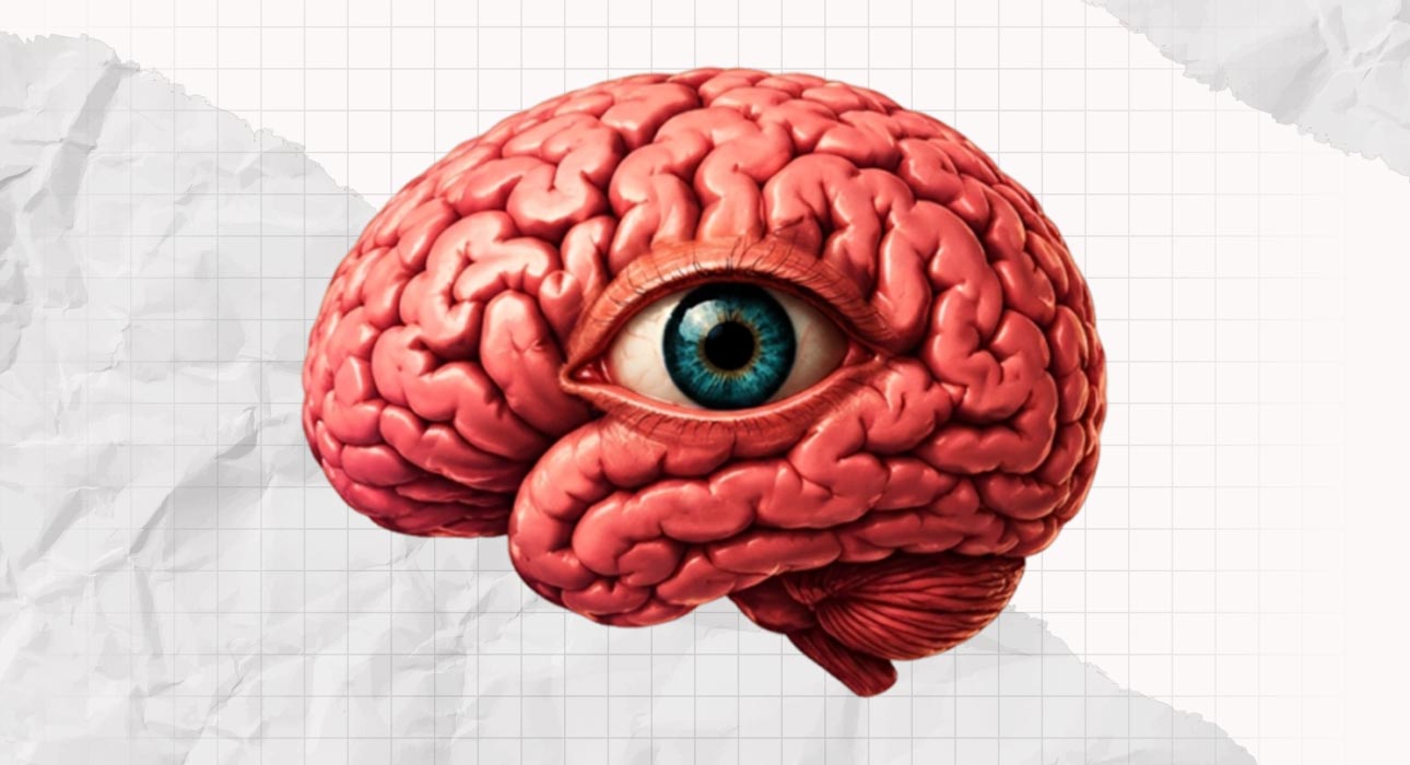
The associative visual cortex then takes the task of matching the image with information from memory to identify the object, giving it meaning. A lesion in the associative visual cortex causes visual agnosia (Álvarez & Masjuan, 2016). But the cerebral location is not enough to understand it. What needs to be discussed as well is what cognitive process helps the vision to make it into a mental image? The answer to that is two processes: perception and association.
Read More: Cerebral Cortex: The most important part of human brain
Two Cognitive Processes That Form What We See
Visual agnosia can be understood by looking into the two types of agnosia related to the two processes of mental images from inside the brain. The cognitive processes of perception and association. Perception processes the form, size and volume of an object; it helps the person to see in a three-dimensional way (Álvarez & Masjuan, 2016). With apperceptive agnosia, the person cannot differentiate a straight line from a curved line. For example, this type of agnosia gives the person the same degree of disability as someone blind.
For example, the patient described by Miller (2018) is called patient D.F. She had a form of visual perception agnosia, and couldn’t copy forms of any object presented to her, even if she could distinguish colours or sequences. The patient presented problems with visuospatial (mentally recognising space between objects) and a poor shape perspective that also affected her recognition of faces and shapes. Association: This is the process in which the brain associates the perceived object with something seen in the past. Patients with associative visual agnosia can draw, differentiate the object from others, but cannot recognise it further (Álvarez & Masjuan, 2016).
Color Without Color
Inside the agnosia spectrum, there is a particular type that relates to how the colour is processed in what is seen. Such as simultagnosia or apperceptive agnosia. Achromatopsia. Achromatopsia is a disorder in which the person is unable to differentiate colours (Álvarez & Masjuan, 2016). The prevalence in the general population is that 1 of 30,000 people has it. This form of agnosia causes impaired colour discrimination, reduced visual clarity, and photophobia (extreme sensitivity to light) (Genead et al., 2011). The cause is a lesion in the bilateral ventral occipito-temporal region (Álvarez & Masjuan, 2016).
These patients can describe the world as a grey world. Clinicians may consider this a minor disability. Nevertheless, according to Álvarez & Masjuan (2016), patients describe extremely unpleasant situations, and it can also affect their everyday life. For example, one patient described saying that they couldn’t stand the colour grey on people’s skin and food and had to close their eyes to eat also began to avoid touching and social interactions (Álvarez & Masjuan, 2016).
Also, achromatopsia is different from other agnosias that have colour as the focal point. For example, colour agnosia, where the patient has a reduced knowledge of colour but no difficulties perceiving them, and colour anomia, where there is difficulty naming colours but no difficulties with perceiving them or knowing the names of colours (Martinaud, 2017).
Another characteristic of achromatopsia is its association with cerebral necrosis (dead tissue) in the posterior region of the brain. Researchers have observed this condition in patients following trauma, encephalitis, and neurodegenerative diseases, while achromatopsia rarely occurs on its own. Also, it can be related commonly to alexia, prosopagnosia and others (Álvarez & Masjuan, 2016).
Read More: Delirium vs. Dementia: Risks of Misdiagnosis and Psychological Consequence
Unable to Recognise Things Around
To better understand the syndromes that can occur alongside achromatopsia, let us briefly examine them. Another recognised subtype of agnosia is:
- Prosopagnosia: Inability to recognise faces, also related to other types of agnosia, rarely seen alone in patients.
- Pure alexia: Also known as “word blindness”, present by an impairment in word reading but no difficulties in oral language and an ability to write spontaneously.
- Dorsal simultagnosia: Patients with this syndrome can recognise most objects but cannot process more than one at a time (Martinaud, 2017).
Are We Aware of What We See and How to Move?
Agnosia cases are unique because trauma or brain damage can affect each person differently, resulting in distinct symptoms and syndromes. From the way the lesion happens to how the person is going to react to that lesion and how the brain of that person is going to react. With the studies of agnosia, a question was on the minds of scientists: how does agnosia affect the visual field of a person?
Milner (2012) goes back to that question by studying two cases: a 70-year-old man and the already-known patient, D.F., and the process of seeing an object is not seeing, and that’s all. No, what Milner says is that in order to move towards an object, the person has full awareness of the movement of the arm, the target object, the shape of the object to pick it up instead of another object, as well as the closest object and so on. All of that process actually involves two areas of the brain that interact during all of that process: the dorsal stream and the ventral stream. Two interconnected neural pathways.
Read More: Predictive Processing: Is the Brain Really a Prediction Machine?
Actual Limitations and Future Researchs
Milner, in order to do his research, used not only perceptual and visual tests on the patients but also used brain scans. This was in order to locate the areas that interact or do not during this process. Behrmann (2025) discusses that while neuro images help with diagnoses and the study of visual agnosia, there needs to be a consideration of other ways of studying those cases. A mix of the study of neural profiles and behavioural observation can be the future of study in the field of agnosia. The use of FMRI’s, 2d and 3d studies and tests, and more precise scans can help locate the most exact position of the patient’s lesion.
Conclusions
The visual process of recognition and perception begins with the retina seeing an object, but to fully understand and recognise an object, it goes through several areas of the brain. Each one of them contributes to the process of an image into a thought where the person recognises the object, names it and remembers its function and, depending on that, makes the decision to reach for it that also involves the visuals of the mechanics of the hand and its surroundings. However, when trauma damages an area of the brain, the person may develop a form of agnosia.
A person with a head injury may develop any of the subclasses of agnosia. The ability to process information differentiates one condition from another and can become impaired. For example, when colour perception is impaired, a person may develop achromatopsia, which can occur alongside another syndrome and affect their daily life, even when clinicians classify it as a minor disability. And the spectrum of agnosia can be present in several ways, such as not recognising faces or places.
References +
- Álvarez, R., & Masjuan, J. (2016). Visual agnosia. Revista Clínica Española (English Edition), 216(2), 85-91.
- Behrmann, M. (2025). Visual agnosia in the era of behavioural and neural investigations. Neuropsychologia, 109299.
- Biran, I., & Coslett, H. B. (2003). Visual agnosia. Current neurology and neuroscience reports, 3(6), 508-512.
- Genead, M. A., Fishman, G. A., Rha, J., Dubis, A. M., Bonci, D. M. O., Dubra, A., … & Carroll, J. (2011). Photoreceptor structure and function in patients with congenital achromatopsia. Investigative ophthalmology & visual science, 52(10), 7298-7308.
- Martinaud, O.(2017). Visual agnosia and focal brain injury. Revue Neurologique, (), S0035378717304940–. doi:10.1016/j.neurol.2017.07.009
- Mccarthy, R. A., & Warrington, E. K. (1986). Visual associative agnosia: a clinico-anatomical study of a single case. Journal of Neurology, Neurosurgery & Psychiatry, 49(11), 1233-1240.
- Milner, A. D. (2012). Is visual processing in the dorsal stream accessible to consciousness? Proceedings of the Royal Society B: Biological Sciences, 279(1737), 2289.
- Milner, A. David. (2018). [Handbook of Clinical Neurology] The Parietal Lobe Volume 151 || Perceptual deficits of object identification: apperceptive agnosia. , (), 269–286. doi:10.1016/B978- 0-444-63622-5.00013-9