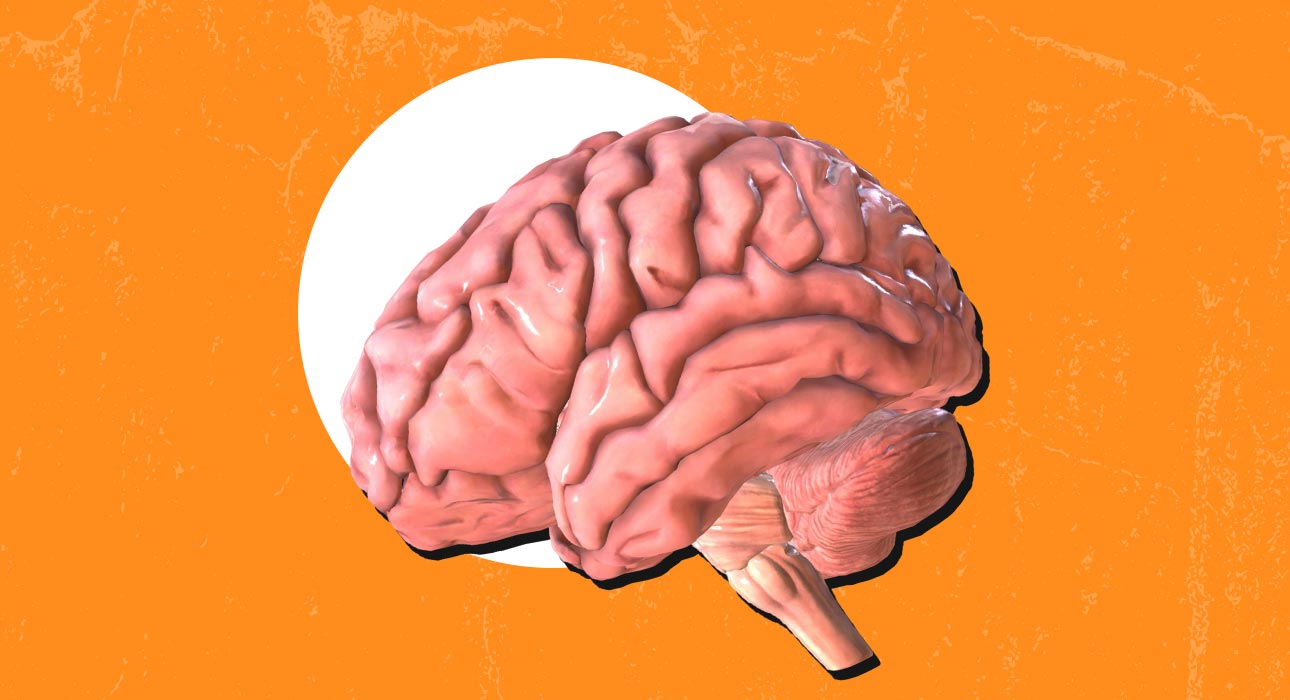
Chronic stress is more than feeling overwhelmed. It is a state that stays with us for a time and can actually change our brains (McEwen & Morrison, 2018). When we are stressed for months or years, our body continues to use the pituitary-adrenal axis, which releases cortisol, the main stress hormone. We need cortisol to survive for a time, but if the levels stay high for too long, it can start to change the brain.
New research using MRI machines shows that chronic stress is associated with brain thinning in the prefrontal cortex (Savic, 2015; Liu et al., 2015). The prefrontal cortex is the part of the brain that helps us make decisions, control our emotions, and behave properly. This research also supports the idea that stress hormones can hurt our brain cells over time, which is called the neurotoxicity model.
To put it simply, chronic stress does not just make us feel bad. It can actually change the brain itself. Chronic stress can make these changes without us even realising it. The brain is affected by stress, and this is what we need to remember about chronic stress.
Read More: The Psychology of Everyday Decisions: How We Make Decisions Without Realising It
Structural MRI Evidence of Prefrontal Cortical Thinning
1. Occupational and Everyday Chronic Stress
One of the most obvious human examples of stress-induced cortical thinning is found in research involving people suffering from chronic occupational stress and exhaustion syndrome (a condition in which the person is exposed to chronic psychosocial stress) (Savic, 2015). The results of the structural MRI analysis are as follows:
- Cortical thinning in the prefrontal areas, including the lateral prefrontal cortex, was associated with the severity of stress.
- Partial reversibility of prefrontal cortical thickness following interventions for stress coping.
2. Mood Disorders & Cortisol Links
In the episode of major depressive disorder, people who have not taken any drugs before have a problem where their body makes too much of a certain chemical. This is often linked to a part of the brain that’s thinner than it should be. Importantly, when people with depressive disorder have more of a certain chemical called cortisol in their blood, the outside part of the front of their brain is thinner. This means that people with major depressive disorder have cortisol in their blood, and the front part of their brain becomes thinner, which is a major problem for people with major depressive disorder.
3. Childhood Stress and Cortical Thickness in Adulthood
Research on people over time shows that stress that happens in childhood can cause problems later on (Monninger et al., 2019). This kind of stress can lead to Reduced cortical thickness in areas in the front part of the brain in young adults. These thin areas in the brain are connected to feeling depressed in life, which means that stress when we are young can change the brain and cause problems that last a long time, especially with the brain’s structure, and this is all related to chronic early life stress and its effects on the brain.
Read More: When Nostalgia Hurts: Understanding Childhood Trauma and Neglect
4. Other Stress-Related Conditions
The result of research done in trauma-exposed individuals with chronic post-traumatic stress disorder (PTSD) shows:
- Thinning of the cortex in the prefrontal cortex and superior frontal cortex (Wrocklage et al., 2017)
- Associations between cortical thinning and maladaptive coping styles and resilience profiles in chronic PTSD.
Altogether, these human MRI studies reveal the same conclusion: chronic stress, whether of a psychosocial, affective, or trauma-related nature, is associated with reduced cortical thickness in the prefrontal cortex.
Mechanistic Models: Glucocorticoid Neurotoxicity and Structural Brain Changes
Glucocorticoid Impact on Neurons
The glucocorticoid neurotoxicity model says that when glucocorticoids, like cortisol, are elevated for a time, they can hurt the brain. This happens in ways.
- The branches of the neurons in the brain, called dendrites, get smaller and have fewer points where they connect with other neurons.
- The growth of neurons and the framework that holds them together is slowed down, which makes it harder for the brain to change and adapt.
- The glucocorticoid receptors in the brain get overactive, which can lead to too much stress on the brain cells when it happens all the time.
The glucocorticoid neurotoxicity model and its effects have been studied a lot in animals, and people are finding out that it is true, for humans too, because the areas of the brain that have the glucocorticoid receptors are also the areas that get the thinnest. (McEwen & Morrison, 2018).
Cortical Regions with High GR Expression
The prefrontal cortex shows increased concentrations of glucocorticoids (GRs), which:
- Provides stress-related feedback but also makes these regions vulnerable when cortisol remains high.
- Show an inverse relationship between regional receptor expression profiles and cortical thickness distributions across the lifespan.
This anatomical receptor is one of the key reasons why the prefrontal cortex is a vulnerable region to the structural effects of stress.
Cellular Pathways
At the cellular level, the increased exposure to glucocorticoids affects:
- Microglial activation and synaptic pruning result in the loss of functional synapses
- Decreases in synaptic scaffolding proteins (such as PSD-95) result in loss of structural integrity.
- Changes in glutamatergic neurotransmission disrupt the structural integrity of the cortex.
Major Depression and Cortical Thinning
When we look at people with Major Depressive Disorder who have hypercortisolemia, which means their body is always producing much cortisol (Liu et al., 2015).
- We use a kind of scan called MRI morphometry, and it shows that the lateral orbitofrontal cortex gets thinner.
- The amount it gets thinner is related to how much cortisol is in their blood, which is what we would expect if cortisol is really bad for the brain.
Major Depressive Disorder patients, with hypercortisolemia, are an example of how being stressed out all the time can actually change the brain in ways.
Read More: The Connection Between Emotional Stress, Depression, and Cardiovascular Health
Implications
These findings are important for understanding how stress that lasts over time affects us. Stress that lasts a time contributes to:
- Cognitive issues, like trouble with executive function, are connected to the prefrontal part of the brain getting thinner.
- Trouble in controlling our emotions because the prefrontal cortex plays a role in managing the limbic structures that deal with emotions.
- Being more likely to get conditions like depression, PTSD and stress-related exhaustion states.
Reversibility and Plasticity
Some studies that go on for a time have found that, when people are stressed, the outside part of their brain gets a little thinner. A positive note is that this thinning can be stopped or even reversed if people learn better ways to deal with stress, like talking to someone or going to therapy. (McEwen & Morrison, 2018)
This means that the brain is good at changing and adapting, so even if it gets a little damaged from stress, it can still get better again. Stress affects the brain. This is what these studies on the brain show that stress and the brain are connected and that the brain can change because of stress, but it can also change back with things like therapy and better coping with stress.
Conclusion
Stress that goes on for a period of time can affect the brain. It can shrink the part of the brain called the prefrontal cortex. Work-related stress can cause this in people. They are sad or depressed, or because they have been through a traumatic event. When we analyse the images of the brain, we can see that stress is the reason for this to happen. When we feel stressed, our body releases a hormone called cortisol. If the release of cortisol happens often, it can damage the brain cells and the connections between them.
Read More: How to Handle Work Stress and Avoid Leaving Your Job?
References +
Liu, X., Kakeda, S., Watanabe, K., et al. (2015). Relationship between cortical thickness and serum cortisol levels in drug-naïve, first-episode patients with major depressive disorder: A surface-based morphometric study. Depression and Anxiety, 32(9), 702–708.
Moreno, G. L., Bruss, J., & Denburg, N. L. (2017). Increased perceived stress is related to decreased prefrontal cortex volumes among older adults. Journal of Clinical and Experimental Neuropsychology, 39(4), 313–325.
Long-term impact of early childhood stress on orbitofrontal cortical thickness (2019). Cerebral Cortex.
McEwen, B. S., & Morrison, J. H. (2018). The effects of chronic stress on the human brain: From neurotoxicity, to vulnerability, to opportunity. Frontiers in Neuroendocrinology, 49, 91–105.
Wong, T. P., et al. (2019). Corticosteroids and regional variations in thickness of the human cerebral cortex across the lifespan. Cerebral Cortex, 30(2), 575–588.
Preprints on GC-induced synaptic deficits and mood disorder progression (2025).
Savic, I. (2015). Structural changes of the brain in stress-related exhaustion disorder.
Cerebral Cortex, 28(3), 894–906 ∙ Monninger, M., et al. (2019). Long-term impact of early life stress on orbitofrontal cortical thickness. Cerebral Cortex, 30(3), 1307–1316.